


Blindness changes the brain differently than previously thought
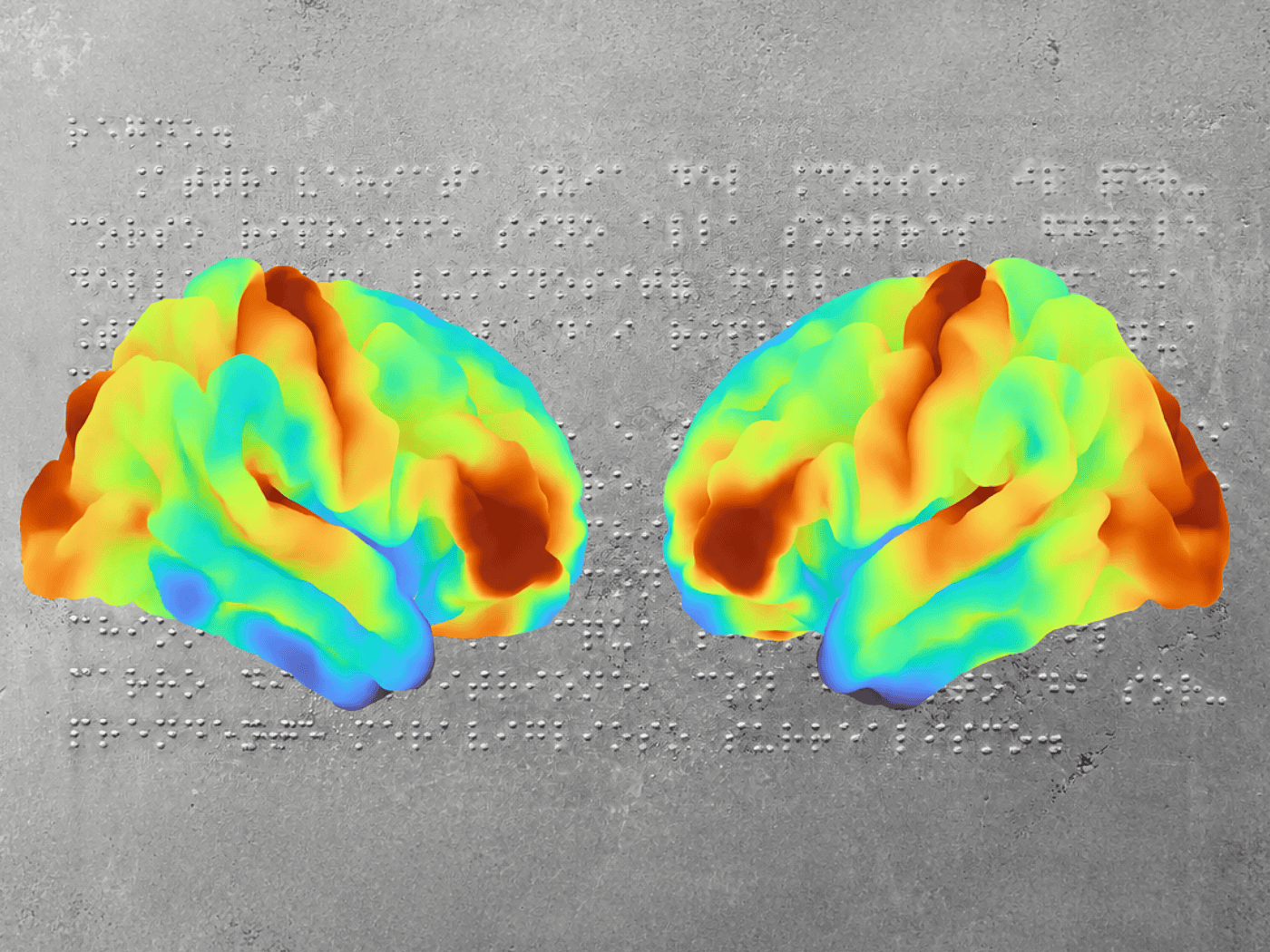
Every day, we rely on our eyes without even thinking about it. But what happens in the brain of a person who has never been able to see? A German-Polish research team has investigated this question. Their findings challenge a previously widely accepted explanation for why the brain develops differently in people with blindness. The study involved Jagiellonian University in Kraków and the Max Planck Institute for Human Cognitive and Brain Sciences in Leipzig.
The brain undergoes significant changes during the first years of life. Initially, it forms more connections between nerve cells than it will later need. It gradually breaks down connections that are rarely used. Experts call this process “pruning.” At the same time, myelination takes place. In this process, a layer of fat envelops the nerve fibers, similar to the insulation on an electrical cable. This allows signals to be transmitted more quickly.
The answer lies in myelin
For years, it has been noted that the visual cortex – the part of the brain that processes visual impressions – appears thicker in people born blind than in sighted people. The conventional explanation to date has been that, without visual input, fewer superfluous neural connections are pruned. To test this, 24 people from Poland who had been blind since birth traveled to Leipzig and underwent MRI scans of their brains. They were compared with sighted control subjects.
Using particularly high-resolution MRI scans, the researchers were able to examine the brain with a level of detail that had previously been virtually impossible. This initially confirmed a well-known observation: the visual cortex appeared thicker in blind individuals than in sighted people. However, the cause was different than previously assumed.
The brain tissue in that area was surrounded by less myelin, a protective fatty layer that accelerates the transmission of nerve signals. The researchers found no evidence, however, that the natural breakdown of redundant neural connections was disrupted.
Different, not impaired
“Reduced myelination can also affect how the boundary between gray and white matter appears in MRI scans,” explains lead author Anna-Lena Stroh. “As a result, the cerebral cortex may appear thicker in MRI measurements.” The researchers also found similar changes in the white matter, which connects different regions of the brain. In areas important for hearing or touch, however, no such differences were observed.
The brains of blind people therefore do not function any worse than those of sighted people. “The visual cortex in blind people is not impaired – it’s just organized differently,” says Marcin Szwed of Jagiellonian University in Kraków. In blindness, it takes on other tasks, such as language processing, working memory, and cognitive control. The researchers’ work thus demonstrates how flexibly the brain can adapt to the absence of visual input.
Original publication:
Anna-Lena Stroh et al., Congenital blindness reduces myelination in the human visual cortex. Sci. Adv. 12, eaec2348 (2026).